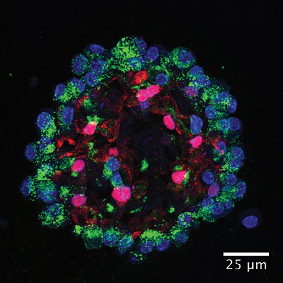

A lung is not just a collection of airway cells. It is a living tissue where structural cells and immune cells work together, and that interaction can be difficult to reproduce in a laboratory dish. A Stanford Medicine team supported by the US National Institutes of Health has reported a human lung organoid model that keeps several of those components together long enough to study how local immune responses work.
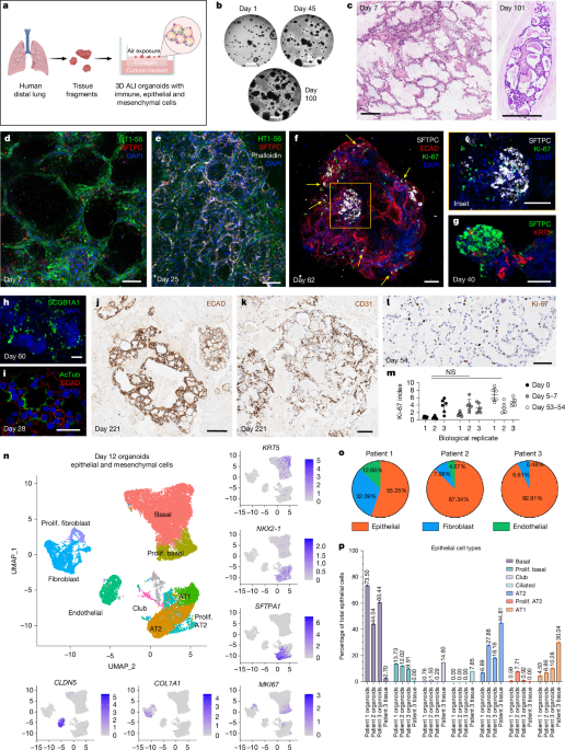
The researchers started with intact distal lung tissue from more than 220 volunteer donors and placed fragments into a three-dimensional air-liquid culture system. The model maintained key lung structures and immune cell populations for up to several months. That duration matters because a short-lived sample can show an immediate reaction but may fail to reveal how resident immune cells persist and respond over time.
The model responded to a virus
To test whether immune cells in the organoids were functioning, the team exposed them to SARS-CoV-2. The model showed general inflammatory reactions and virus-specific adaptive responses, including T cells reacting to the virus. The researchers also pre-stimulated some organoids with viral fragments before infection. In this laboratory setup, the immune cells responded more effectively and subsequent infection was reduced.
That result gives scientists a new way to examine immunity in the lung itself. An organoid can preserve tissue relationships that are lost when cells are separated and grown individually. It may let researchers ask how local immune memory changes with infection, age or disease, and how an intervention affects the lung’s first line of defence.
A research model is not a treatment
The experiment took place in cultured tissue. It did not test a new vaccine in volunteers, and it does not show that a person can be protected by inhaling a viral fragment. The NIH describes the system as a platform for research. It may help answer questions that animal models cannot, but researchers will need to reproduce the findings and test the model across different donors and conditions.
There are practical limits as well. Lung tissue culture is technically demanding, and a laboratory model cannot fully reproduce the circulation, nervous system and other organs that shape human health. The model’s strongest value may be as one tool in a wider research programme, used alongside clinical studies and other laboratory methods.
Even with those boundaries, keeping immune cells alive in human lung tissue for months is a meaningful advance. It gives researchers a closer view of the tissue where respiratory infections begin and offers a more direct way to study local immunity. If the platform holds up across future studies, it could help scientists test ideas about respiratory vaccines and disease without mistaking a promising dish-based result for a proven medical intervention.
Sources
Image credits
Cover image: ZEISS, human distal lung organoid reference image. In-article image: Rathkey et al., Nature, 2026, Figure 1.
The conversation
0 comments
Join the discussion with a thoughtful response.